GST 융합단백질의 발현과 정제
원핵생물의 세포를 이용하여 단백질을 발현시킬 경우, 대장균 내에서 외래성의 유전자를 대량 발현시켜, 이것을 조제하는 것이 많다고 생각된다. 대장균에 대량 발현된 단백질을 조제 할 때 기본적인 수법의 예를 들면, 그 때의 몇개의 문제점을 지적하고자 한다.
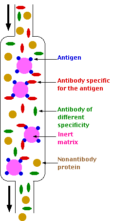
대장균으로 목적단백질을 발현시켰을 때, cDNA을 연결하는 벡터에 무엇을 선택할지가 문제이다. 목적단백질만을 순수하게 얻고 싶은지, 또는 다른 적당한 단백질과의 융합단백질로서 목적에 맞는지가 문제이다. 각각에 적당한 벡터가 시판되고 있는지, 또는 구축한 자로부터 구할 수 있는 경우가 많다. 융합단백질은 발현한 대장균의 추출액으로부터 발현벡터에 code되어진 배열에 대한 affinity chromatography에 의하여 1단계에서 정제할 수 있다라는 이점이 있다. 그러나, 고차구조 해석 등과 같은 목적을 위해서는 다음에 이 여분을 protease처리로 제거할 필요가 있다.
여기서는 일반적으로 연구실에서 먼저 검토한 발현벡터로 glutathione-S-transferase (GST) 와의 융합단백질을 만드는 pGEX 벡터를 이용한 경험을 예로 설명한다.
이 벡터는, tag promoter (Ptag) 를 갖고, lacIq 지배하에 있는 isopropyl - b - D - thiogalactoside (IPTG) 로 발현의 유도가 걸리게 된다. 이러한 벡터에는 여러 종류의 multi cloning site (MCS) 를 갖는 것이 사용하기 쉽다. 발현한 융합단백질도 glutathione을 결합시킨 Sepharose수지로 용이하게 정제할 수 있다. 또, GST 유전자와 MCS Sepharose의 간에 특이성이 높은 protease 인식부위가 삽입되어 있다. 결합단백질로서 발현 정제한 후, 적절한 potease를 이용함으로서 GST domain (26kDa) 을 제거하는데 유용하지만, 사전에 목적단백질 분자내에 protease 인식 배열이 존재하지 않는 것을 확인할 필요가 있다.
본 항에서는 융합단백질의 발현 및 정제와 융합단백질로부터 GST domain을 잘라내어 목적단백질을 얻는 수법에 관해서 알아 보고자 한다.


Purification of GST-protein from E.Coli
Lysis buffer
-100mM Tris-HCl, pH 7.6, 100mM NaCl, 1 mM EDTA, 1 mg/ml lysozyme, 1 mM DTT, 1 ㎍/ml of pepstatin, leupeptin
Wash buffer
-50mM Tris-HCl, pH 7.6, 150mM NaCl, 1% NP-40,1mM DTT, 1 ㎍/ml of pepstatin, leupeptin, 10% Glycerol
1. 50ml tube에 5ml LB+antibiotics 에 seeding 하여 37 도에서 overnight culture
2. 250ml 의 LB+antibiotics 첨가하여 1 시간 culture at 37 도 -> before induction : 400ul
3. final 0.5-1mM IPTG 첨가하여 4-6 시간 culture at 37 도 -> after induction : 400ul
4. 2500rpm 에서 20분 centrifugation [harvest]
5. 상층액을 완전히 제거한 후에 Lysis buffer8ml 에 다음을 첨가한 후 첨가할 것.
+ 7mM DTT + 100ug/ml lysozyme + 1ul of aprotinin and leupeptin (stock: 10mg/ml)
6. 잘 섞어서 ice 에서 20 분간 방치.
7. sonication -> after sonication: 40ul
8. 80 ul of 10% dodexycholate was added and incubated at RT for 20 min.
9. 25 ul of 10 mg/ml DNAse was added, incubated at RT for 20 min.
10. 13000rpm for 30 min at 4oC, supernatant를 취함.
11. 300 ul 의 G-bead 를 Lysis 1ml buffer 로 equilibration (X5).
12. step 10에서 얻어진 sup 을 이용하여 step 11에서 equilibration 해 놓은 G-bead binding for 2 hr at 4 oC.
13. centrifuge 하여 G-bead 를 wash buffer로 8 회 washing.
14. The beads were resuspended to a 50% solution in the processing wash buffer with 20% glycerol
15. Frozen in aliquots at -80 oC.
GST pull-down
Lysis Buffer
-50 mM Tris, pH 7.6, 150 mM NaCl, 10 mM EDTA, 10 mM sodium pyrophosphate, 10 mM NaF, 1mM sodium orthovadate, 10 ㎍/ml of pepstatin, leupeptin, and AEBSF each, 1% NP-40.
Wash Buffer
-20 mM Tris, pH 7.6, 150 mM NaCl, 10 mM EDTA, 10% glycerol, 5 mM NaF, 1mM sodium orthovadate, 1 ㎍/ml of pepstatin, leupeptin, and AEBSF each, 0.1% NP-40.
1. 293 cells were transfected on 100-mm plates with 10 ㎍ of plasmid.
2. At 48 hr transfection, the cells were washed with ice cold PBS and lysed with 1 ml lysis buffer.
3. Incubation for 20 min at 4℃, the cell lysates were cleared through centrifugation at maximal speed for 10 min.
4. The supernatant was collected and incubated with pre-washed beads with 1~2 ㎍ of GST-protein for 2 hr at 4℃.
5. The beads were washed four times with wash buffer.
6. The beads were boiled with 100 ㎕ of 1X SDS-PAGE loading buffer, and the proteins were by SDS-PAGE and analyzed by Western blotting.